The best diagnostics are achieved when high-resolution
images such as those from TRACE (0.5)
are combined with spectroscopic data
(CDS resolution @ 4).
There are very few simultaneous CDS and TRACE multifilter
AR observations.
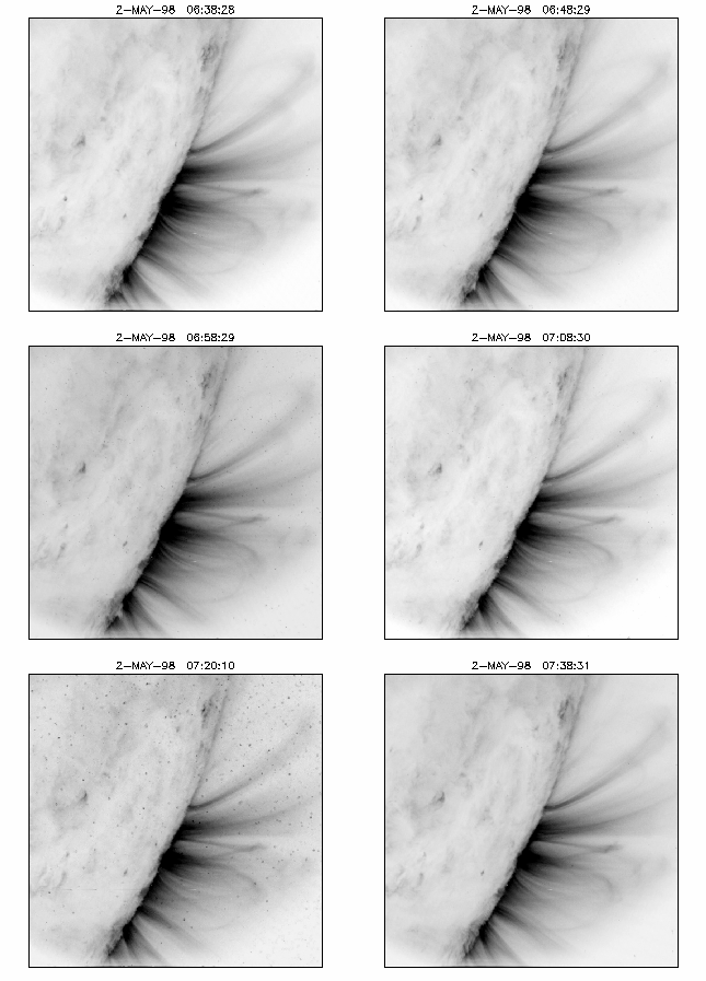
Figure 13: Sequence of TRACE 173 Å negative images of the
active region.
Many loops are visible.
The images are plotted with the same intensity scale, and
show that the loops were stationary during the whole
period of the CDS observation.
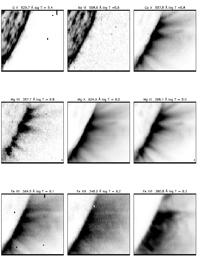
Figure 14: Monochromatic images (negative) of the NIS observation.
The loops are only seen in upper transition region
lines (Mg VII, Ca X, Mg IX) emitted in the range
Log T = 5.8-6.0, a clear indication of their
isothermality.
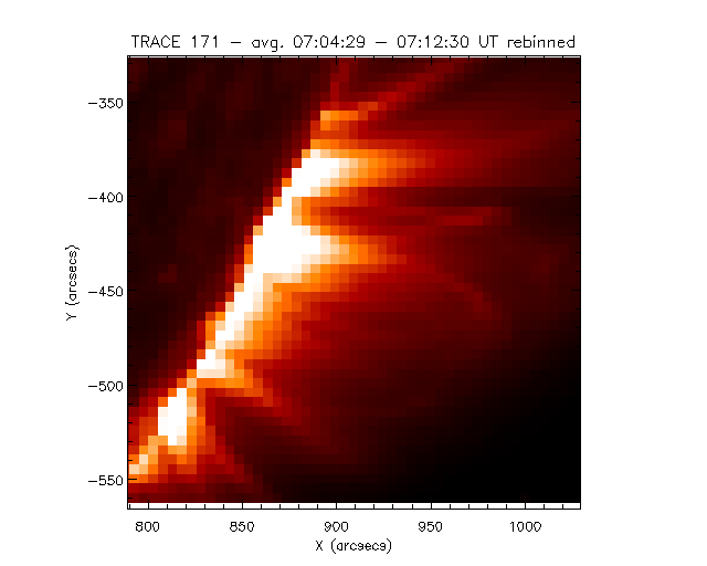
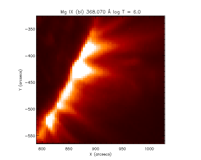
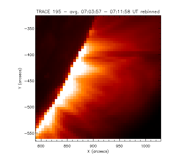
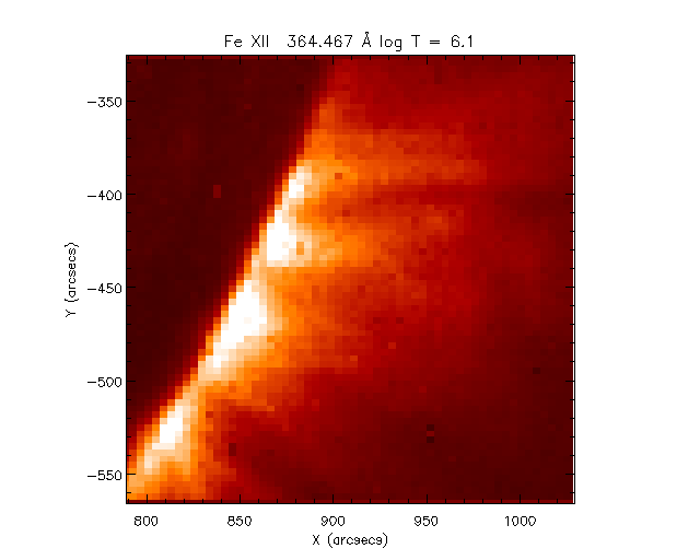
Figure 15: From top to bottom:
TRACE 173 and 195 Å negative images of the
active region;
the same images rebinned by a factor of 10
(each pixel size=5);
the CDS images in Mg IX and Fe XII