| Department of Applied Mathematics and Theoretical Physics |
Stephanie S.M.H. Höhn - Senior Research AssociateMechanics of MorphogenesisGoldstein group SRATrinity Hall PDRA Equality & Diversity Researcher Representative (DAMTP) CamAWiSE (Cambridge Association for Women in Science & Engineering) Steering Group Member, Former Deputy-Chair I believe that a diverse team and environment are crucial for the success of scientific research. Research How do cells generate the forces that shape our tissues and organs? In developing embryos, cells move and change their shape in an astoundingly coordinated way. We need to understand both the underlying mechanics and genetic regulation, as errors in this self-organisation can lead to birth defects. And when these cell behaviours are triggered in adult diseases they can even cause cancer. Many tissues, including the primal gut, the neural tube and our retina, are formed through Cell Sheet Folding.
I combine advanced imaging, experiments and computational modelling to reveal the underlying biophysical and mechano-chemical mechanisms.
The Mechanics of Epithelial Folding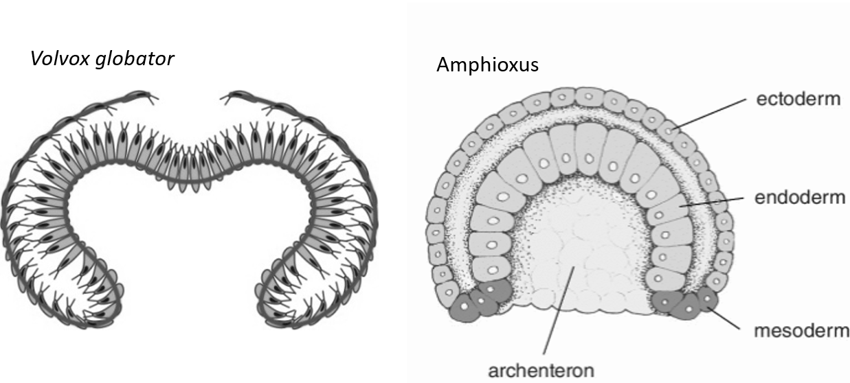 I am using a comparative approach to reveal the overarching mechanical principles governing invagination (inward folding) of cellular monolayers. Early embryos of the simple Chordate Amphioxus (the closest relative of vertebrates) and the green microalga Volvox are uniquely suited to study the dynamics and mechanics of cell sheet folding: They consist of spherical cellular monolayers that are accessible to direct measurements and perturbations of mechanical properties, fluorescent live imaging and computational modelling. Understanding the folding of cellular monolayers will serve as the groundwork to untangle the mechanisms driving more complicated events in invertebrates (e.g. echinoderms, cnidarians, ascidians) and vertebrates.
This work is funded by independet grants awarded by the Cambridge Centre for Physical Biology and the Society for Developmental Biology.
I am using a comparative approach to reveal the overarching mechanical principles governing invagination (inward folding) of cellular monolayers. Early embryos of the simple Chordate Amphioxus (the closest relative of vertebrates) and the green microalga Volvox are uniquely suited to study the dynamics and mechanics of cell sheet folding: They consist of spherical cellular monolayers that are accessible to direct measurements and perturbations of mechanical properties, fluorescent live imaging and computational modelling. Understanding the folding of cellular monolayers will serve as the groundwork to untangle the mechanisms driving more complicated events in invertebrates (e.g. echinoderms, cnidarians, ascidians) and vertebrates.
This work is funded by independet grants awarded by the Cambridge Centre for Physical Biology and the Society for Developmental Biology.
Inversion, a gastrulation-like process in Volvox globator; selective plane illumination microscopy (SPIM). ECM Expansion and Cell Packing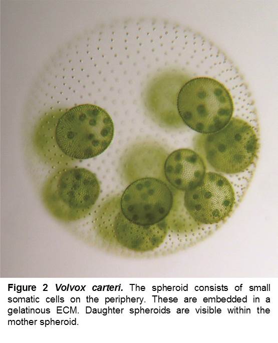 The microalga Volvox carteri is used as a model to study how the positioning of cells emerges from a combination of cell division patterns and ECM synthesis. Juvenile V. carteri spheroids consist of a spherical cellular monolayer, embedded in ECM. With increasing ECM production the spheroid grows, the cells move apart and end up in a polygonal pattern. We seek to unravel how this cell positioning is regulated. Through a combination of advanced imaging and computational simulations we were able to show that the cellular organisation in both Volvox and lab-grown snowflake yeast obeys a maximum entropy law.
Volvox cartericonsists of ca. one thousand small somatic cells and few reproductive cells (gonidia) (fig. 2). Each somatic cell possesses two flagella which enable the organism to swim and perform phototaxis. All cells are embedded in a gelatinous extracellular matrix (ECM) that is secreted by the cells. In order to allow for coordinated swimming, the flagellated cells need to be positioned regularly within the ECM. Very little is known about the self-organisation of the correct cellular patterns in Volvox [5].
I am using cell tracking techniques and time-lapse microscopy to reveal the underlying mechanisms of cell positioning in the developing spheroids.
Research expertiseMolecular cell biology (mutant strain generation, gene expression profiling), imaging (light sheet microscopy, CLSM, TEM, SEM), biophysical experiments (micropipette-aspiration, micro-manipulation, laser ablation) and theory (shell theory, visco-elasticity), and computational methods (GUI-based modelling platforms, Matlab, Comsol). I am using custom numerical models in collaboration to explore the mechano-chemical signalling processes giving rise to tissue deformations.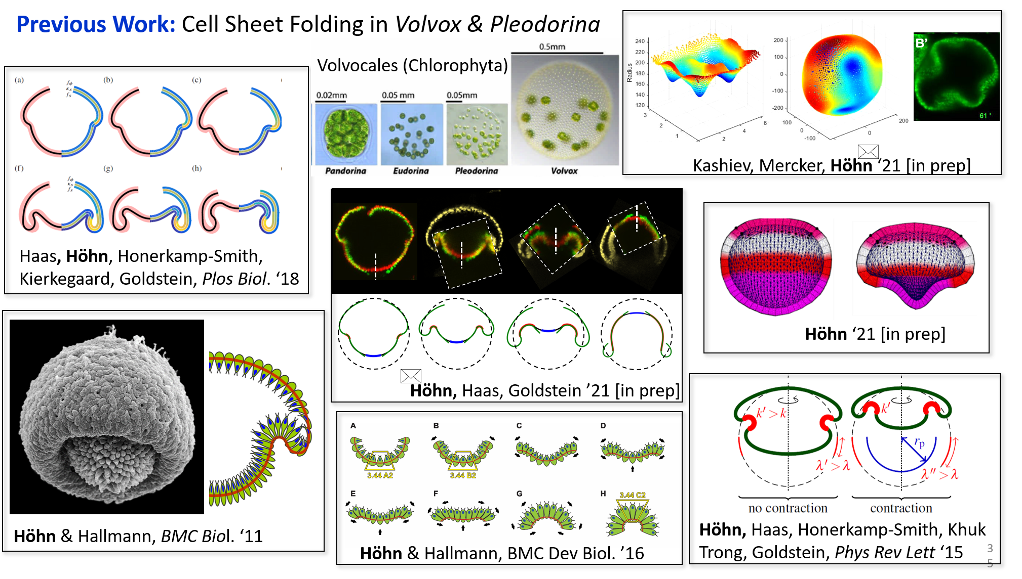
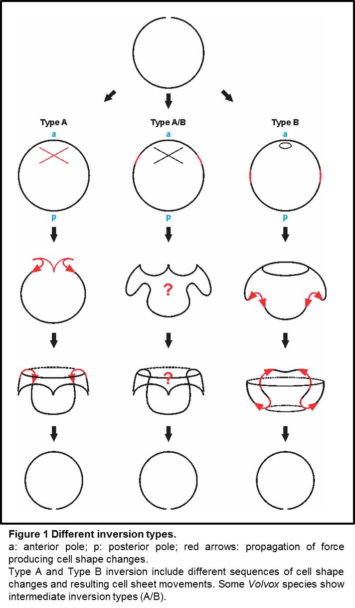
Cell sheet folding is a common morphogenetic process during the development of multicellular organisms. Spheroidal green algae of the genus Volvox are uniquely suited as simple model systems for studying the basic principles of epithelial folding. Volvox embryos consist of a cellular monolayer which turns itself inside-out to achieve its adult configuration; this process is called inversion.
Different Volvox species have different tactics to turn their embryos inside out (fig. 1). These different types of inversion involve varying sequences of global shape changes driven by local active cell shape changes.
I am seaking to reveal the relation between local cellular changes and global deformations.
Society membershipsRoyal Microscopy SocietyBiophysical Society Society for Developmental Biology Previous workType B inversion in Volvox globatorI did my PhD research with Prof. Armin Hallmann in the Department of Cellular and Developmental Biology of Plants at the University of Bielefeld, on the embryonic type B inversion in Volvox globator. 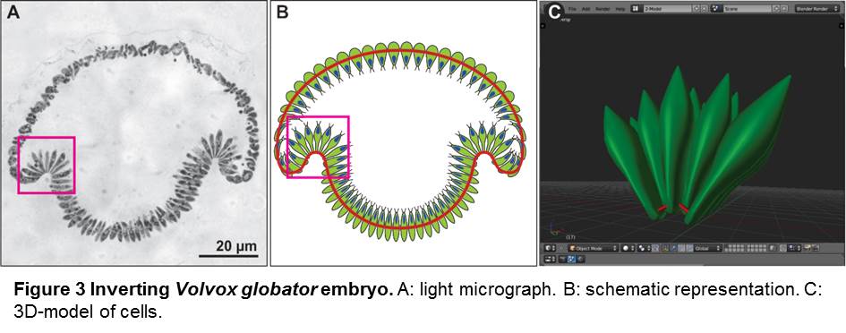
|